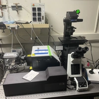
Advanced Imaging Facility

The imaging facility is managed by Dr. Andrey Malkovskiy, who can be reached at amalkovskiy@carnegiescience.edu.

The imaging facility currently has 13 major instruments, two dissecting microscopes, 3 instruments in reserve (two DMI6000, one Horiba Labram HR800 Raman instrument), 4 major software packages for image analysis or acquisition, and associated supporting hardware. There are two dedicated computers for image analysis, one has remote access. Remote access via RemotePC software is available for most major instruments to users by request. Training and Agendo reservation system registration is required for all instruments.
Leica SP8 Point Scanning Confocal Microscope with white light laser (WLL)
The SP8 is a state of the art point scanning confocal microscope. It produces the best out of focus rejection of all the instruments in the facility (most "confocal": highest contrast, best axial resolution) and has the most flexible array of excitation and emission bands, due to WLL wide-range illumination and two additional lasers. However, it is not the most sensitive instrument for small numbers of chromophores and live cell imaging. The SP8 has tools for performing photobleaching and photoactivation experiments, optical anisotropy and FLIM/FRET capabilities with very high time domain resolution (4 ps). Additional software from Leica is installed as well: Navigator, Lightning deconvolution.
Description of Leica SP8:
Excitation lines: 405, 440, 470-670 nm tunable wavelength and frequency pulsed WLL.
Emission bands: continuously customizable over three detector channels with AOBS system
Detectors: 2 HyD high-sensitivity confocal, PMT confocal, transmitted light PMT
Objectives: 63X Oil n.a.=1.4, 63x Glycerin n.a.=1.3, 63x water n.a.=1.2, 40x oil n.a.=1.25, 20x multi-immersion n.a.=0.7, 20x long-working air 0.6 n.a.
Software: Leica Navigator, Leica Lightning deconvolutio
Modules: FLIM, FRAP, optical anisotropy, WLL pulse-picker
Sign-up: Carnegie.agendoscience.com
Requirements for use: Training required. Contact Andrey Malkovskiy.
Room: 408
Example:

Spinning Disk Confocal Microscope 1
The spinning disk instruments have less out of focus rejection than does the SP8, but they have superior sensitivity cameras for extended imaging of dim subjects in living cells. Both systems feature high speed imaging acquisition and piezo-driven stage focusing, multiple laser lines for excitation, high speed excitation selection, shuttering and attenuation by AOTF, and detection by back-thinned, high QE EMCCD cameras. The SD-1 Olympus system also utilizes proprietary silicone oil objectives for superior refractive index matching to biological tissues. Incubation system for microscopes is installed. Light-sheet capability is optional and can be enabled on this system.
Available excitation lines: 442, 488, 514, 561 nm
Excitation/emission pairs: 442/cyan plus 514/yellow, 488/green plus 561/red (see scope manual for exact specs)
Stand: Olympus IX83 with automated focus control, motorized focus, objective turret and dichroic carousel, YOKOGAWA spinning-disk CSU-W1
Objectives: 100x Oil n.a.=1.4, 100x Sil Oil n.a.=1.35, 40x Sil Oil n.a.=1.25, 20x air n.a.=0.7
Camera: Andor iXon Life. 512x512, QE greater than 90%, on chip electron multiplication
Auxiliary focusing available: MadCity Labs piezo focusing stage.
Multichannel imaging: Dualview image splitter for GFP/RFP or CFP/YFP
Incubation chamber: TOKAI HIT incubation system
Software: SlideBook
Requirements for use: Training required.
Room: 401
Example:
Spinning Disk Confocal Microscope 2
Similar to SD-1, this SD-2 system has superior camera sensitivity, but also has high-power lasers for FRAP photobleaching. The system features high speed imaging acquisition, multiple laser lines for excitation, high speed excitation selection, shuttering and attenuation by 3i programmable AOTF, and detection by back-thinned, high QE EMCCD camera.
System 2 also has a 405 nm excitation line for near-UV excitation of chromophores such as DAPI, a camera with improved signal to noise characteristics, and a fully motorized stage for repeated visitation of multiple sample locations and for automated image tiling.
Available excitation lines: 405, 442, 488, 514, 561 nm.
Excitation/emmission pairs: 442/cyan plus 514/yellow, 488/green plus 561/red, 405/green/red (see scope manual for exact specs).
Stand: Leica DMI6000 with Adaptive Focus Control, motorized focus, objective turret, dichroic carousel, field stop, condenser, YOKOGAWA spinning-disk CSU-X1
Objectives: 100x Oil n.a.=1.4, 63x Oil n.a.=1.4, 63x glycerol n.a.=1.3, 63x water n.a.=1.2, 20x Multi-immersion n.a.=0.7, 20x air n.a.=0.7
Camera: Photometrics Evolve EMCCD 512x512.
Auxiliary focusing available: MadCity Labs piezo focusing stage.
Incubation chamber: Oko Touch from Okolabs with Lauda ECO RE 415 ethylene glycol cooling
Software: SlideBook.
Location: Room 402
Example:

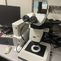
Fluorescence Stereoscope Nikon SMZ18
Ideal for fluorescence or brightfield whole seedling or colony screening. Equipped with motorized Z stand and automatic filter cube wheel. Can perform timelapse and Z stack imaging.
Objective: 1X macro
Filtercubes: ECFP, TagRFP, GFP-bandpass, GFP longpass, custom chloroplast
Camera: RGB color Nikon DS-Ri2
Light source: Nikon Sola
Software: Nikon NIS
Example:
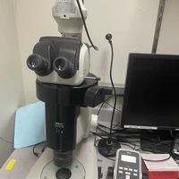
Fluorescence Stereoscope Leica M165FC
Ideal for fluorescence or brightfield whole seedling or colony screening
Objectives: 0.6X macro, 1.6X macro
Filtercubes: DSR ET, GFP2, GFP3
Camera: RGB color Leica DSC320
Light source: Lumen Dynamics X-Cite Series 120PC Q
Software: Leica LAS
Example:
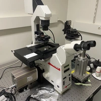
Fluorescence upright Nikon
For fluorescence imaging where inverted imaging is not possible or convenient. Has differential imaging contrast (DIC) options
Stand: Nikon ECLIPSE E6000
Objectives: 60x oil n.a.=1.4, 60x water n.a.=1.2, 40x air n.a.=0.75, 20x air n.a.=0.75, 10x air n.a.=0.45
Filtercubes: GFP, RFP, DAPI
Camera: RGB color Luminera Infinity 3
Software: Infinity Capture
Fluorescence upright Leica
For fluorescence imaging where inverted imaging is not possible or convenient. Has differential imaging contrast (DIC) and Phase options
Stand: Leica DMRB
Objectives: 100x oil n.a.=1.3, 40x air n.a.=0.7, 20x air n.a.=0.5, 10x air n.a.=0.3
Filtercubes: A, I3, N2.1
Camera: Nikon D7000
Software: Nikon Camera Pro
Vertical macroimaging system
Ideal for fluorescence or brightfield whole seedling or colony screening with very powerful 8x extra-long working distance objective with variable aperture
Stand: Custom, Thorlabs optics with kinematic mounts, ASI motorized XYZ stage, Sutter filter wheel
Objective: Leica 8x M PL Apo MACRO with 48 mm working distance n.a.=0.22
Camera: Roper Quantem 512SC
Software: MicroManager
Example:
Horizontal macroimaging system
Roper CoolSNAP EZ camera, ASI motorized XYZ stage, Sutter filter wheel for whole seedling or colony screening.
Software: MicroManager
Example:
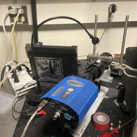
Ratiometric epifluorescence imaging system
Ideal for analysis of FRET sensors in mammalian cells (HEK293T cells, HepG2 cells), in arabidopsis roots or leaf slices.
Custom assembled system: Sutter light source Lambda DG4, millisecond switching of excitation wavelengths, equipped with Chroma ET/sputtered excitation filters, liquid light guide,
Stand: Leica DMIRE2
Objectives: 20x n.a.=0.7
Camera: Roper Quantem 512SC
Vacucell perfusion chamber for mammalian cells, World precision perfusion chamber for plants, Automate computerized perfusion control system, Sutter emission filter wheel, Roper Dualview image splitter
Software: MicroManager
Example:
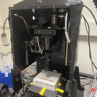
FEI Quanta 200 Environmental SEM
The Quanta 200 is a very flexible instrument with XYZ, rotation and tilt that allows wet and living specimens to be imaged without special preparation, along with classic high-resolution imaging. It features a cooling stage and 5 detectors: backscatter, ET, Large Field Detector, Gaseous Secondary Electron Detector 500 and 1000 microns
Sign-up: Agendo
Room 410
Example:

Sample preparation:
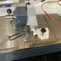
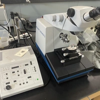
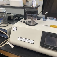
Thin sample preparation for fluorescence and TEM imaging
Leica RM2025, Leica RM2065, Vibratome 1500 for thicker sections
Leica Ultracut S for sub-micron sections
Denton Vacuum DCP-1 supercritical drying with liquid CO2
Denton Vacuum Desk IV sputtering with gold targets.
Microtomes Example:
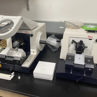
Leica Ultracut S Example:
Denton Desk IV sputtering example:
Glass knife maker example